Laboratorios de Diagnóstico Oftalmológico
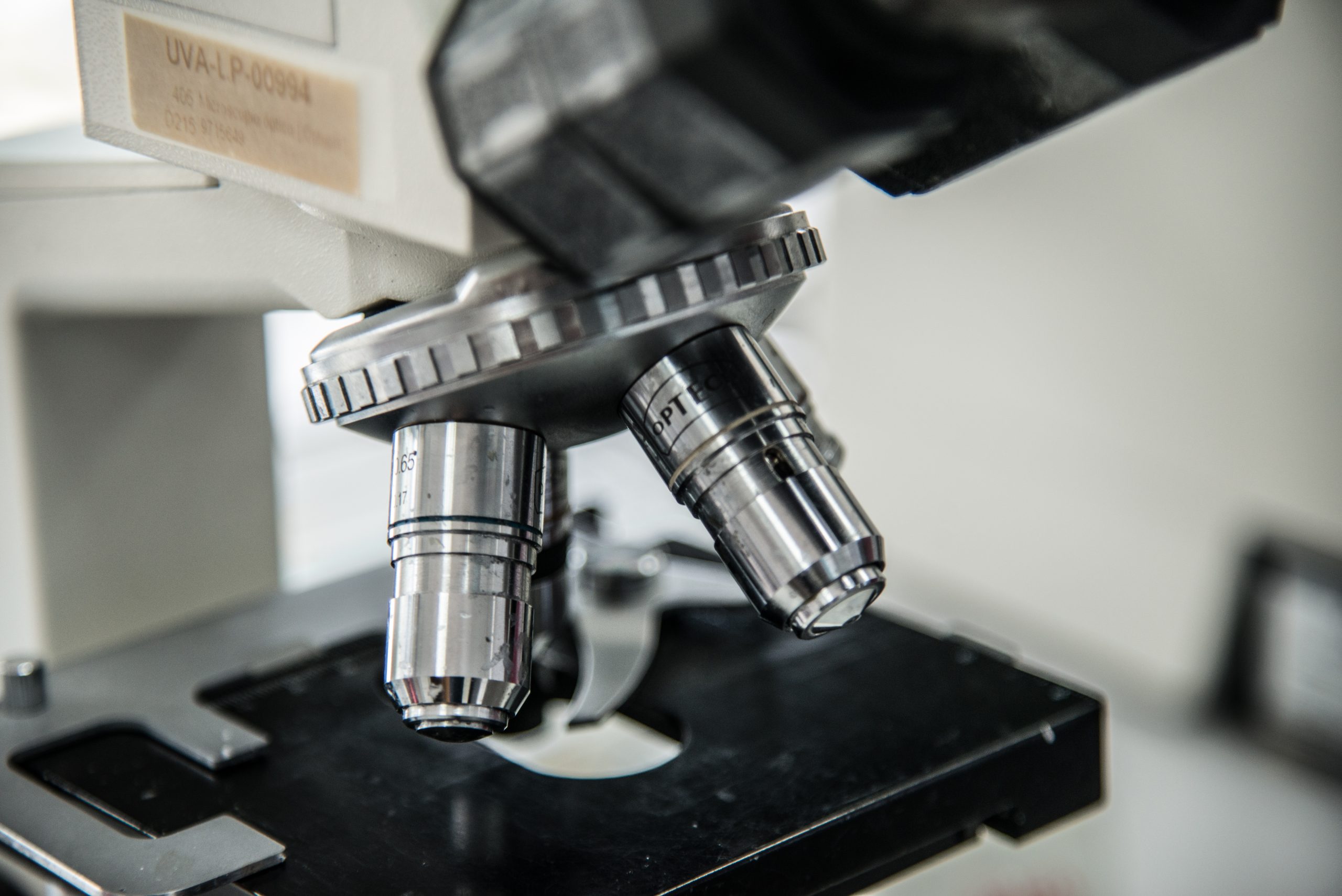
Laboratorio de Patología Ocular (LPO)
Ofrecemos todo el material disponible en nuestro archivo (bloques de parafina y preparaciones histológicas) para actividades docentes y de investigación, lo que supone más de 7000 muestras almacenadas (6,226 informatizadas 31/12/2021).
Realizamos el procesado y la evaluación histológica de muestras correspondientes a proyectos de investigación, tanto humanas como de animales de experimentación.
Teniendo en cuenta la legislación vigente, el Instituto Universitario de Oftalmobiología Aplicada (IOBA) ha desarrollado y aplica un Procedimiento de Gestión de muestras biológicas de origen humano con fines de investigación biomédica.
Publicaciones
Purpose: We evaluated the expression of the neural markers, neuron-specific enolase, and synaptophysin, as a tool to confirm the diagnosis of retinoblastoma (RB) in undifferentiated and advanced tumors. Additionally, we determined whether the extent of RB-associated protein (pRb) expression is helpful in assessing the prognosis in RB patients.
Methods: Conventional whole tissue section and tissue microarray immunohistochemistry for neuron-specific enolase, synaptophysin, and pRb were carried out in a series of 22 RBs. Results: Neuron-specific enolase and synaptophysin were expressed in 75%–100% of the tumor cells, and the staining intensity was strong. Two RBs expressed pRb in 75%–100% of the tumor cells, also with strong staining intensity. Concordance between the immunohistochemical outcomes for whole tissue staining and tissue microarray staining was 76.2% for neuron-specific enolase, 85.7% for synaptophysin, and 80.0% for pRb.
Conclusion: Neuron-specific enolase and synaptophysin have the potential to be useful markers for the diagnosis of RBs. Extensive and strong pRb staining is not associated with less aggressive tumor behavior according to the pathologic classification of RBs.
Keywords: retinoblastoma, immunohistochemistry, neuron-specific enolase, synaptophysin, pRb
Purpose: The purpose of this study was to characterize the central epithelial thickness (CET) of penetrating keratoplasty corneal specimens obtained from patients with keratoconus (KC) and correlate the histological patterns with their clinical history.
Methods: Ex vivo histological imaging was performed to measure CET and total corneal thickness (TCT) in 56 patients with KC. Microscopic slides from penetrating keratoplasty corneal specimens, stained with hematoxylin and eosin were evaluated using bright field microscopy.
CET and TCT were measured, and morphological features were studied. Clinical history regarding duration of KC prior to surgery and length of and tolerance to contact lens wear were compared and analyzed.
Results: The microscopic slides of all patients available for follow up (n = 48) were analyzed and CET and TCT were measured. The histological evaluation revealed 3 distinctive epithelial patterns. Pattern 1 with central hypertrophic and hydropic changes (n = 19) measured 70.89 ± 25.88 m in CET and 308.63 ± 100.74 m in TCT; Pattern 2 (n = 14) had not changed,
similar to normal epithelium CET and TCT measuring 36.5 ± 7.02 m and 260.14 ± 87.93 m respectively. Pattern 3 (n = 15) demonstrated thinner central epithelium characterized by atrophy and focal hydropic changes measuring 19.93 ± 4.60 m and 268.00 ± 79.39 m in CET and TCT respectively (all p < 0.0001). The presence of Pattern 2 characterized by similar to normal CET was correlated with the duration of the condition (R = 0.600, p = 0.030). There was a significant difference in the length of CL wear comparing those with patterns 1 and 2 versus 3 (least no. of CL years) (p = 0.05 and p = 0.33 respectivelly).
Purpose. &e purpose of this study was to confirm the presence of specific patterns of epithelial response in corneal buttons from keratoconus patients.
Methods. &is was a retrospective and descriptive study. 90 penetrating keratoplasty specimens obtained from patients diagnosed with keratoconus were evaluated using bright-field microscopy. Morphologically identifiable characteristics including epithelial cell density and epithelial thickness were analyzed on hematoxylin and eosin- (H&E-) and periodic acid of Schiff- (PAS-) stained slides.
Results. &ree distinctive patterns of epithelial alteration of the central cornea were established. Pattern 3, in which the central epithelium was as thick as peripheral epithelium, was the commonest (44.4%), followed by the pattern 2, defined as central epithelium thinner than periphery epithelium (38.9%), and the uncommonest pattern was number 1, with central epithelium thicker than the periphery (16.7%). Conclusions. &ree distinctive histologic patterns that could potentially have a diagnostic and prognostic value in keratoconus patients were found.
Profesionales
Antonio López García
Técnico de Laboratorio
Antonio López García
Técnico de Laboratorio
Dr. Juan Cuevas Álvarez
Doctor especialista en Anatomía Patológica
Dr. Juan Cuevas Álvarez
Doctor especialista en Anatomía Patológica
Dra. Elena Pérez
Doctora especialista en Anatomía Patológica
Dra. Elena Pérez
Doctora especialista en Anatomía Patológica
Especialidad en Anatomía Patológica en el Hospital 12 de octubre (2000-2004)
Facultativa especialista en el Hospital Universitario Río Hortega de Valladolid (Actualmente)
Área de especialidades: Oftalmopatología, Dermapatología, Uropatología, Patología de Pulmón y Ginecología
Laboratorios/unidades de investigación clínica: Laboratorio de Patología Ocular /LPO)

Dra. Marina Gándara Cortés
Doctora especialista en Anatomía Patológica
Dra. Marina Gándara Cortés
Doctora especialista en Anatomía Patológica